1. Hasegawa, A. and Mori, W.: Morphometry
of the human pineal gland; Relationship to the adrenal cortex. Acta
Pathol Jpn, 30:407-411, 1980) (pdf 750KB available).
Abstract: To investigate the role of the pineal gland in the endocrine system, we examined 26 autopsy cases dying from non-neoplastic diseases. The study was carried out on the hypothetic basis of the possible influences of the pineal gland on the pituitary-adrenal axis, which we believe to be existing also in man (BS, BS, BS, humankind -- politically correct in 1997, Akio). Measurements on the pineal gland revealed that the weight of the gland was directly proportional to the volume (r=0.94; P less than 0.001) and also to the nuclear density of the parenchyme (r=0.65; P less than 0.01). Then, the interrelationship between the weight of the pineal gland and the thickness of the adrenal cortex was determined (r=-0.56; P less than 0.05). The pineal gland was significantly larger in weight in the prednisolone treated patients (166 +/- 66 mg. [+/-SD]) than in the others (97 +/- 52 mg; P less than 0.05). We conclude that the functional antagonism exists between the pineal gland and the adrenal cortex in man, although the mechanism involved has not been clarified yet.
2. Hasegawa, A. and Mori, W.: Pituitary-adrenal axis in pinealoma. Acta Pathol Jpn, 32: 925-931, 1982 (pdf 243KB available).
Abstract: Autopsy series of 19 patients with pineal and anterior third ventricle (suprasellar) tumors were analyzed with regard to shift in pituitary-adrenal axis. Germinomas formed the most common subgroup in this series (79%). Metastatic tumor growth in the pituitary gland caused adrenal atrophy in 4 cases. Some patients with pineal neoplasms had larger adrenals in weight compared with age- and sex-matched controls of Japanese. In three patients with sexual precocity, adrenal weight was larger. It is concluded that clinical importance exists in anterior pituitary insufficiency with adrenal atrophy, but hyperfunction of the hypophyseoadrenal system might occur during the course of the disease.
3. Hasegawa, A., Izumiyama, N., Ohtsubo, K., and Mori, W.: The increase of the nuclear lobulation of the
pinealocytes in aging mice; Electron-microscopic karyometric study. Jpn J
Aerospace and Environ Med 24; 23-28, 1987 (pdf 1.1MB available).
Abstract: The age-dependent changes of the nuclei of the mice pinealocytes were studied electron microscopically. The morphometrical results showed that (1) the perimeter of the pinealocyte nuclei increased during 5 to 8 months of age (3 to 5 & 8 months; P less than 0.05), then gradually decreased to 24 months with advancing age (5 & 8 to 24 months; P less than 0.05), and (2) the mean number of the nuclear segments per cell increased according to age (r = 0.793, N = 22, P less than 0.0001). These findings may reflect the age-associated changes in the cytokinetic and metabolic activity of the pinealocyte nuclei.
4. Hasegawa, A., Ohtsubo, K., and Mori,
W.: Pineal gland in old age; Quantitative and qualitative morphological
study of 167 human autopsy cases. Brain Research, 409: 343-349, 1987 (pdf 1.1MB available).
Abstract: Age-related changes of human pineal gland were studied morphologically on 168 autopsy cases. Pineal weight in the formalin-fixed condition was 99 +/- 56 mg in males (n = 88, mean age 78 years) and 91 +/- 41 mg in females (n = 80, mean age 79 years), which showed no sexual difference (P = 0.290). There was no correlation between pineal weight and age of the patient (r = 0.0678, P = 0.191, n = 168). The pineal volume calculated as a spheroid was directly proportional to the weight (r = 0.904, n = 167, P = 0.000). A significant correlation existed between pineal weight and the degree of calcification (r = 0.231, P = 0.002, n = 156). The density of the pineals decreased with age (r = -0.164, P = 0.017, n = 167). The degree of calcification and cyst formation did not increase with age, but the grade of cyst formation was related to the pineal volume (P = 0.0002). Some pineals of the patients over 90 years old showed no calcification and appeared indistinguishable from the ones of the younger subjects. The weight and volume of the pineals of the patients with hypertension was appreciably greater than those of the non-hypertensive patients. These results indicate that human pineals do not necessarily degenerate progressively after involution.
5. Hasegawa, A., Ohtsubo, K., Izumiyama,
N., and Shimada, H.: Ultrastructural study of the human pineal gland in
aged patients including a centenarian. Acta Pathol Jpn, 40; 30-40, 1990 (pdf 1.3MB available).
Abstract: An ultrastructural study of human pineal glands obtained at autopsy from 7 patients older than 70 years was conducted in order to clarify the functional anatomy of the pineal in the aged. By light microscopy, the pineal glands from aged patients were parenchymatous and almost indistinguishable from those of younger controls. Electron microscopy of the pineal parenchymal cells revealed deep nuclear indentations, synaptic ribbons and ribbon fields, Golgi apparatus, lipofuscin granules and microtubular sheaves in all subjects, cilia with a 9 + 0 pattern in a few, and lamellated structures suggestive of the outer segment of photoreceptor cells very rarely. Microtubules were numerous in the cytoplasmic processes and bulbous endings. Fibrous astrocytes located between the pinealocytes showed long and thin cytoplasmic processes containing numerous glial filaments. Two types of nerve bouton were present in the pineal parenchyma, one of which contained clear vesicles forming synapse-like contacts with pinealocytes. There were no significant age-related changes in these features in a qualitative comparison with pineal glands from 5 adult patients younger than 70 years. These findings indicate that even in advanced age, the human pineal gland maintains some functions, such as intercellular communication and photoreception, in common with the pineal in lower vertebrates.
6. Hasegawa, A., Shimada, H., Izumiyama, N., and Ohtsubo, K.: Paired twisted filaments in human pinealocytes. Acta Pathol Jpn, 41; 265-269,1991 (pdf 5.0MB available).
Abstract: Paired twisted filaments (PTF) have been confirmed and described ultrastructurally in the normal human pineal gland for the first time. The PTF showed a peculiar double helical structure, measuring 12 to 25 nm in maximal helix width with a half-periodicity of 30 to 35 nm (periodicity of the constrictions). Each filament was about 10 nm wide. The PTF formed parallel aggregates in the perikaryotic cytoplasm of the pinealocytes. In 8 of 12 autopsied middle-aged to elderly individuals, the PTF were present in a small proportion of pinealocytes. The identity of the PTF remains unclear in terms of their apparent similarity to the paired helical filaments observed previously in aged rats. However, the age distribution of individuals with PTF suggests that the intermediate filaments of human pinealocytes are more susceptible to early deterioration during aging than those of other cerebral neurons.
1. Mori, W., Machinami, R., Shiga, J.,
Tanaka, K., Hasegawa, A., et al.: A pathological study of fulminant
liver disease. Acta Pathol Jpn, 34:727-742, 1984.
2. Masuda, M., Takano, Y., Iki, M., Makiyama, K., Ikeda, I., Noguchi, S., Hasegawa, A., and Hosaka, M.:
Cyclin-dependent kinase inhibitor p27Kip1 expression in transitional cell carcinoma of renal pelvis and ureter. Cancer Letters, 150; 183-189, 2000.
3. Sugimura, H., Mori, H., Nagura,K., Kiyose, S., Hong, T., Isozaki, M., Igarashi, H., Shinmura, K. Hasegawa, A., Kitayama, Y., Tanioka, F.: Fluorescence in situ hybridization analysis with a tissue microarray - 'FISH and chips' analysis of pathology archives. Pathol International, 60; 543-550, 2010 (pdf 667KB available after registration to Wiley online library).
1. Kitamura, K., Asai, M., Kubo, T.,
Harii, K., and Hasegawa, A.: Mucinous carcinoma of the external
auditory canal; case report. HEAD & NECK, 12:417-420, 1990.
2. Yagishita, A., Nakano, I., Ushioda, T.,
Otsuki, N. and Hasegawa, A.: Acute encephalopathy with bilateral
thalamotegmental involvement in infants and children: Imaging and
pathology findings. Am J Neuroradiol, 16:439-447, 1995.
3. Hasegawa, A.: Fibrolamellar
hepatocellular carcinoma: A report of a resected case with an electron
microscopic and flow cytometric analysis. Pathology International,
46:84-90, 1996 (pdf 2.8MB available).
Abstract: A resected case of fibrolamellar (FLC) and hepatocellular (HCC) combined carcinoma arising in a non cirrhotic liver of a 29 year old female is reported, including results of the preoperative percutaneous aspiration biopsy, which suggested FLC, and postoperative electron microscopic and flow cytometric analysis. Sections of the resected massive tumor of the left lobe of the liver showed hepatocellular carcinoma accompanying the fibrolamellar carcinoma element which was composed of tumor cells with eosinophilic granular cytoplasm and unique cytoplasmic vacuoles (pale bodies). Lamellar fibrosis was present in the stroma, while no macroscopic central scar was demonstrated. Electron microscopy showed typical features of FLC and Row cytometric DNA analysis indicated diploid DNA pattern with a low proliferation rate. A common HCC element with trabecular structure also existed at the periphery of the tumor. No apparent etiologic agent for the development of hepatic neoplasm was indicated in the history of this patient. She had been without recurrence for about 3 years after extended left lobectomy, when local recurrence was revealed. The recurrence has been treated with chemoembolization and percutaneous ethanol infusion several times up till the present. This case reconfirms the importance of the pathological diagnosis of FLC to promote surgical intervention.
4. Hasegawa, A. and Iwasaki, T.: Rhomboid secretion granules in a juxtaglomerular cell tumour of the kidney. Br J Urol, 79:296-297, 1997.
5. Hasegawa, A.: Juxtaglomerular cell tumor of the kidney:
A case report with electron microscopic and flow cytometric
investigation. Ultrastructural Pathology (Taylor & Francis)
21:201-208, 1997.
Abstract: A juxtaglomerular cell tumor (JGCT) was found in the right kidney of a 17-year-old female who had suffered from a headache of 1-year duration and hypertension. CT and US were diagnostic in association with determination of plasma renin activity. Light and electron microscopic, immunohistochemical, and flow cytometric investigation of the resected tumor confirmed the diagnosis of JGCT with demonstration of ultrastructural features and distribution pattern of characteristic secretion granules of various sizes and shapes, coexistence of mast cells within the tumor (JGCT cells/mast cells = 10), and perivascular architecture.
6. Hara, Y., Tajiri, T., Matsuura, K, Nagamoto, A., Jinza, S, and Hasegawa, A.: Malignant fibrous histiocytoma of the spermatic cord: A case report. Nishinihon J. Urol, 66:304-309, 2004.
7. Hara, Y., Hattori, Y., Matsuura, K.,
Hasegawa, A., Jinza, S., and Nagamoto, A.: Adrenal cortical carcinoma:
A case report. Nishinihon J. Urol, 66:593-599, 2004.
8. Hara, Y., Hattori, Y., Matsuura, K.,
Hasegawa, A., Tajiri, T., Jinza, S., and Nagamoto, A.: Adult mature
teratoma of the testis: A case report. Nishinihon J. Urol, 66:703-707,
2004.
9. Hara, Y., Tajiri, T., Matsuura, K., and
Hasegawa, A.: Acute scrotum caused by Henoch-Schoenlein purpura. Int.
J. Urol, 11:578-580, 2004.
10. Hara, Y., Hattori, Y., Matsuura, K.,
Jinza, S., Nagamoto, A., Iwamura, M., and Hasegawa, A.: Multilocular
cystic renal cell carcinoma: A case report. Nishinihon J. Urol,
67:499-506, 2005.
11. Hattori, Y., Hara, Y., Matsuura, K.,
and Hasegawa, A.: Granulomatous balanoposthitis after intravesical
Bacillus-Calmette-Guerin instillation therapy. Int. J. Urol,
13:1361-1363, 2006.
12. Maitani, F., Fujimori, S., Hayashi, Y., Hasegawa, A., and Iwasaki, M.:
A case of juvenile primary pulmonary malignant fibrous histiocytoma. Tokai J. Exp. Clin. Med., 35:130-132, 2010.
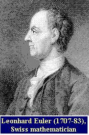
[Review] He was a real genius! It is estimated that an average of 800 pages of new mathematics per year were published over a career of 60 years. And the father of 13 children. What a productivity! By the way, who do you speculate fed them, who cared them, who cleaned their clothes? It was not him. Undoubtedly, Mrs. Euler.
For an example of his genius, he proved in 1734 that a specific infinite series, aka Basel Problem, converges to ¦Ð2 (square of pi; Ludolphsche Zahl)/6. Famous mathematicians had known that it converges to a finite sum, but no one knew "to what sum?". (see Dunham W. The Mathematical Universe. John Wiley and Sons, NY, pp. 51-57, 1994, Paul J. Nahin. An Imaginary Tale: The Story of the Square Root of Minus One. Princeton University Press, New Jersey, pp. 146-149, 1998, and Dunham W. Euler. The Master of Us All. The Mathematical Association of America, 1999)Back to previous Home Page at AOL (Till October 31 2008)
1 + 1/4 + 1/9 + 1/16 + 1/25 + ,,,,+ 1/N2 + ,,,, = ¦Ð (pi)2/6
I could understand the derivation he made by Prof. Dunham's accounts, but I couldn't resist verifing this inference by writing a short program in C (as a Sunday programmer, I love Deitel H. M. and Deitel P.J. C How to Program. Prentice-Hall, NJ, 1992). If you or your friend can use C compiler, I recommend you cut and paste the following program and save it as a file, e.g., "euler.c". Then compile this with "cc -o euler euler.c -lm" or "cc euler.c -lm", when you should never forget "-lm" that links the object code to the math library functions, and run with "euler" or "a.out" that is executable. Or if fortran (f77) is available, which can automatically compile C program as well, "f77 euler.c" is also valid to produce an executable image "a.out".
Note: I have done these largely on Linux, SLS 1.05, softlanding software.
/* Euler's Infinite Series, programmed by Dr. Hasegawa on March 8 1995 */
#include<stdio.h>
#include<math.h>
main()
{
int n;
double s, i;
printf("\n *** Euler's Infinite Series *** \n");
printf(" S = 1 + 1/4 + 1/9 + 1/16 + ,,,, \n\n");
printf("Input N = ");
scanf("%d", &n);
s = 1.0;
for (i = 2; i <= n; ++i){
s = s + pow (1./ i, 2.);
}
printf("\nN = %d", n);
printf("\tS = %.13e\n", s);
printf("\t PI**2/6 = 1.6449340668474e+00\n\n");
}
Voila! If you increase the number N, this series is increasing marginally to ¦Ð (pi)2/6, but really never exceeds it. Brilliance, elegance? I can't select proper word to express my respect for him.
Personally, I am using this program with a stopwatch as a floating-point benchmark for my collection of MPUs including Intel 8086, 80286, 80386, 80486, Pentium, Motorola 68020, Mips R4400 series etc., whose data corresponding to each N (=10x) are stored in a spreadsheet. Just for fun. :)
Back to previous Home Page at geocities.jp web host (Till March 31 2019)
Back to New Home Page (Translocated Sakura server)